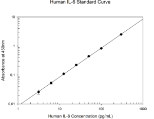
Protein Expression Analysis

Contact Us

ELISA

We have a Synergy 2 plate reader with a red-shifted PMT, a Cytation 3 plate reader containing monochromators that can measure a wide range of wavelengths, and a 405LS Microplate Washer to reduce incomplete or uneven washing. We have extensive experience using ELISAs from multiple vendors and with multiple sample types such as cultured cells, whole blood, fresh tissues, and frozen tissues. Absorbance-based or fluorescence-based ELISA kits can be used to produce a standard curve and accurately quantify the amount of a specific protein in your samples. To obtain the most accurate results, we routinely generate standard curves from protein standards in duplicate or triplicate and quantitates your protein of interest in each sample from triplicate wells.
Human IL-6 Standard Curve

ELISpot / FluoroSpot

Our in vitro services now offers the sandwich enzyme-linked immunospot (ELISpot or FluoroSpot) assay to measure the frequency of cytokine secreting cells in cell suspensions.  This assay allows the secreted cytokine to bind to the coated antibody resulting in a single spot. The CTL S6 Universal analyzer is used to capture quality images and analyze the spots.  Equipped with four optical filters, the CTL analyzer is capable of detecting up to three analytes simultaneously using either colorimetric or fluorometric readouts.  ELISpot or FluoroSpot kits are compatible with blood, lymphoid, and solid tissue.  The samples are routinely performed in duplicate or triplicate to obtain an average to quantify the amount of spots in each sample.

ELISpot

Western blot

In collaboration with our clients we have performed many successful Western blots against a large number of oncology relevant targets. The staff has extensive experience performing Western blots from multiple sample types such as cultured cells, fresh tissues, and frozen tissues. With the Typhoon Imaging System, chemiluminescent, chemifluorescent, and fluorescent secondary antibodies can be detected allowing for the identification of multiple proteins of interest at one time. We also have the ability to quantify the bands on a membrane with ImageQuant software to determine the size and relative intensity of each band for each sample.

Western blot

Flow Cytometry

We have two Attune NxT flow cytometers with four lasers capable of detecting fluorescent compounds in 14 channels. Whether you need to examine the abundance of a specific protein or multiple proteins on the surface or inside cells, our flow cytometry experts together with the Attune NxT can make it happen. Monitor the abundance or localization to/from the plasma membrane of a specific protein over time with treatment or compare the levels of a protein in different cell lines or mouse strains. Combine the detection of a specific protein with any of our other common flow cytometry cell process assays, (below) such as the apoptosis, cell cycle, or cell viability assay to determine if the abundance of the protein changes at different stages of a cell process. We have successfully detected hundreds of different proteins in samples such as cultured cells, blood, tissues, and organs. In blood, we have successfully detected proteins using a no-wash no-lyse assay which reduces the amount of manipulation and allows the analysis of very small volumes of blood.

Flow cytometry

Cytometric bead assays

We also have a Luminex® 100™ System. This system is a flexible analyzer based on the principles of flow cytometry. The system enables you to multiplex (simultaneously measure) up to 100 analytes in a single microplate well, using very small sample sizes. The system delivers fast and cost-effective bioassay results on many assay formats including nucleic acid assays, receptor-ligand assays, immunoassays, and enzymatic assays. Commercial, pre-made, multiplexed plates are available to measure the components of entire cell signaling pathways or cytokines such as the AKT pathway, apoptosis, chemokines, cytokines, inflammatory cytokines, growth factors, and the TH1/TH2 pathways.

Cytometric bead assays

Immunohistochemistry

Analyze your tissue samples with IHC staining to detect abnormal cells such as those found in cancerous tumors. Specific molecular markers are characteristic of particular cellular events such as proliferation or cell death (apoptosis). IHC is also widely used in basic research to understand the distribution and localization of biomarkers and differentially expressed proteins in different parts of a biological tissue. We offer a full suite of in-house histology and IHC services including custom work, and will be adding markers routinely. Our services include:

  • Panels: Cancer, Immuno-Oncology
  • Normal Tissue, Tumor, Xenograft, Cell Line, Validation, Immunofluorescence, Immunocytochemistry, Multiplex
Immunohistochemistry

Fluorescence microscopy

We have an Olympus CKX53F inverted fluorescence microscope to view fluorescent compounds/proteins in live cells and a BioTek Cytation 3 imaging multi-mode reader to view fluorescent compounds/proteins in live or fixed cells. With the use of fluorescence microscopy on live cells, we can quickly determine the percentage of cells containing a fluorescent compound or protein without manipulating the cells. With fixed cells, we can determine the cellular localization of specific proteins within the cells after staining with fluorescently-labeled antibodies and using fluorescent dyes such as DAPI, FM 1-43, Calcein, or DiOC6 to define specific compartments within a cell.

Fluorescence microscopy

Let's start a conversation

Contact Us