Author: Dylan Daniel, PhD, Director, Scientific Development
Date: November 2017
ID8-Luc is a novel syngeneic mouse model developed to treat orthotopic ovarian cancer. Ovarian cancer remains a significant unmet medical need with a relative 5-year survival rate of 17% in patients diagnosed with highly advanced stage IV ovarian cancer.
Ovarian cancer is an unmet medical need and particularly lethal because it is often detected only when disease is quite advanced. In 2017, approximately 22,440 women will be diagnosed with ovarian cancer in the United States, and approximately 14,080 deaths will occur. Combination platinum and anti-mitotic taxane based chemotherapy is the standard of care in the U.S., but while these treatment options are often initially efficacious, relapse and progression usually occur. There is a need for novel therapeutic approaches to treat ovarian cancer.
Based on the recent successes of harnessing the immune system to treat other cancers, drug discovery scientists have explored immunotherapy approaches in ovarian cancer. There are currently upwards of 50 clinical trials testing immunotherapy approaches and combinations in ovarian cancer; however, there are a limited number of syngeneic mouse ovarian cancer models available to enable preclinical pharmacology. Labcorp has developed the ID8 syngeneic mouse cell line model for testing standard therapeutics as well as for immuno-oncology applications. ID8 was derived from C57BL/6 mouse ovarian surface epithelial cells that were transformed by serial passage in vitro.1 Luciferase was expressed in the ID8 cells (ID8-Luc-mCherry-Puro) to enable monitoring of orthotopic (intraperitoneal) tumor growth by bioluminescence imaging (BLI).
The mean tumor growth kinetics (Figure 1A) and individual growth kinetics (Figure 1B) of ID8-Luc-mCh-Puro as monitored by BLI are illustrated. The implantation of 1.0×10⁷ cells per mouse yielded more consistent tumor growth than 5.0×10⁶ cells per mouse in C57BL/6 albino mice. As expected, the signal (photons/sec) is higher in the C57BL/6 albino mice (green line, Figure 1B, group 2) than wild type C57BL/6 mice (red line, Figure 1B) due to a lack of melanin pigment in their skin which absorbs light output from the tumor cells. Representative BLI images of ID8-Luc-mCh-Puro orthotopic disease progression are shown in Figure 1C.
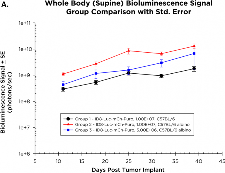
Fig. 1A: Intraperitoneal growth of ID8-luc-mCh-puro ovarian cancer monitored by whole body BLI.
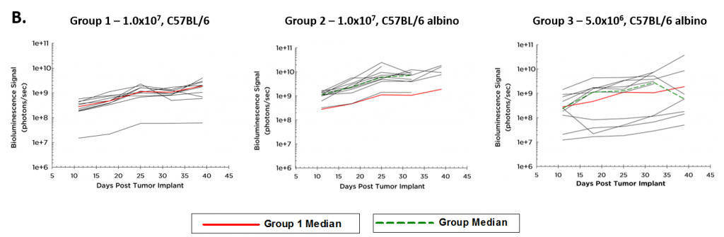
Fig. 1B – Growth Kinetics of Tumor-ID8-Luc-mCh-Puro
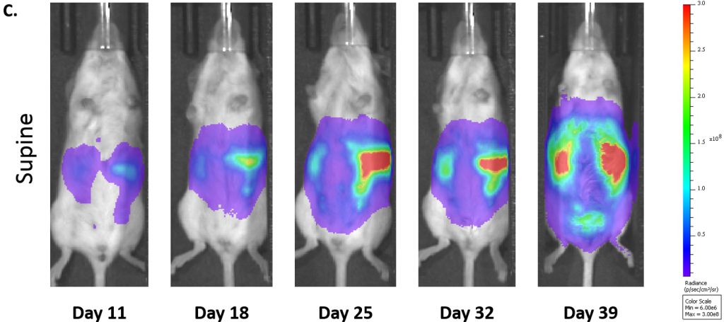
Fig. 1C: Representative BLI Images of ID8-luc-mCh-Puro Ovarian Cancer Progression in C57BL/6 Albino Mice (1.0×10⁷ cells/mouse).
Mice implanted with ID8-Luc-mCh-Puro cells gain weight over the duration of the study (Figure 2) with some mice showing weight loss only at end of study. Disease progression, manifesting as abdominal distention due to ascites formation (Figure 3), is linear with survival of 35-40 days in the C57BL/6 albino mice implanted with 1.0×10⁷ cells.

Fig. 2: Body Weight Change in ID8-Luc-MCh-Puro Model: Mean Body Weight Change by Group with Std. Error.
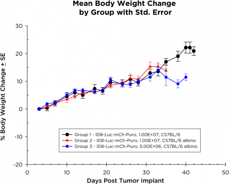
Fig. 3: Survival Kinetics of Mice with Orthotopic ID8-luc-mCh-Puro Ovarian Cancer.
Labcorp performed immune profiling of the ascites from the ID8-Luc-mCh-Puro bearing mice. In Table 1, the immune profiles for total CD45+ leukocytes, CD4+ T cells, CD8+ T cells, Tregs, M-MDSC, G-MDSC, M1 TAM and M2 TAM are shown. While there is considerable heterogeneity in the immune profile between mice, all mice have a robust CD8+ T cell infiltration with favorable CD8+ T cell/Treg ratios. The ID8 ovarian cancer model has been reported to respond to IL-12 immunotherapy which combined with the favorable CD8+ T cell/Treg ratio suggests that the model should support testing of novel immunotherapies.2 Studies to analyze response to immunotherapies as well as standard of care are currently under development.
Table 1: Flow Cytometry Profiling of Immune Cell Subsets in Individual Mouse Ascites from Mice with ID8-Luc-mCh-Puro Ovarian Cancer
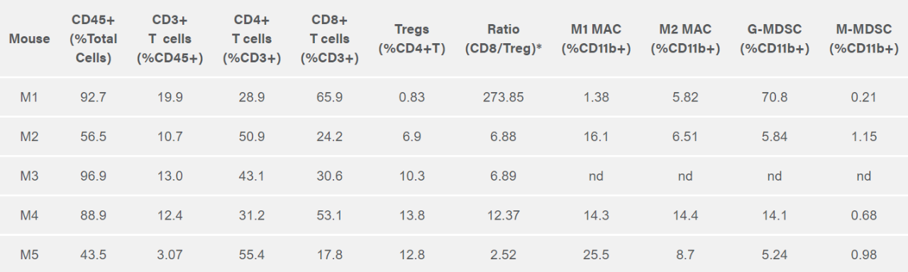
Table 1: Flow Cytometry Profiling of Immune Cell Subsets in Individual Mouse Ascites from Mice with ID8-Luc-mCh-Puro Ovarian Cancer
*CD8/Treg ratio determined based on the absolute counts from the individual CD8 and Treg gates.
nd = not determined
References
Note: Please note that all animal care and use was conducted according to animal welfare regulations in an AAALAC-accredited facility with IACUC protocol review and approval.
Connect
Let's start a conversation
Contact Us
Labcorp is a leading global life sciences company that includes contract research and developmental services to the pharmaceutical, medical technology, crop protection and chemical industries.