Doctor working on a scan

Computed Tomography (CT)

Contact Us

Micro computed tomography (CT) imaging platform draws on expertise in implementing, developing, optimizing, and validating micro-CT imaging paradigms for drug research studies. Our experience in CT-based bone and soft tissue imaging encompasses a number of disease states.

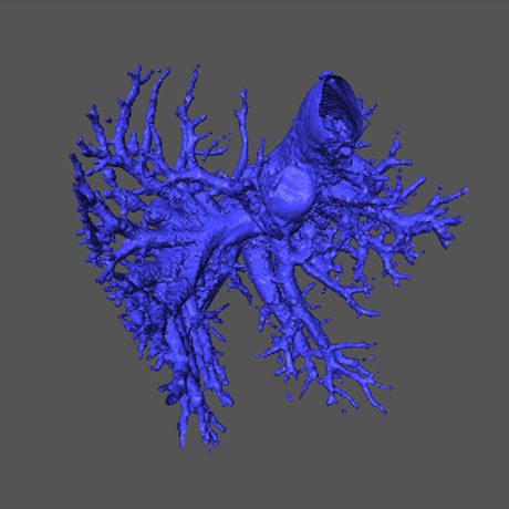
In vivo micro-CT imaging enables high-resolution (to ~50 microns), three-dimensional image generation by rotation of an x-ray source and detector around an imaging subject. Multiple projections are obtained during the rotation and reconstructed to generate the three-dimensional image. The large density differences between bone, soft tissue, and air make them readily distinguished by CT. CT contrast between different soft tissues is generally more subtle. The use of systemically administered, CT contrast agents enables better visualization of the soft tissues, including the vascular system.

Bone Imaging and Metastasis

CT is the preferred, translational technology for the quantification of metastatic and arthritic bone disease. We provide numerous, validated bone-disease models, including several models of bone metastasis and rheumatoid arthritis. Micro-CT can be used to characterize:

  • Bone loss and growth

  • Bone density and microstructure

  • Bone remodeling and calcification

We also have strong expertise in imaging of bone metastasis in validated intracardiac and peritibial models, including imaging of osteolytic and osteoblastic activity, using staged-treatment or prevention paradigms.

Tumor Volume Quantification

Bone Metastasis

We are effectively able to monitor and quantify orthotopic lung tumor growth noninvasively by CT.  We offer three lung models using human tumor cell lines, one PDX lung model, and 17 additional lung tumor cell lines, each of which can be luciferase enabled for evaluation in an orthotopic setting.

Skeletal Phenotyping

Micro-CT provides non-destructive imaging for 3D analysis of bone architecture. Protocols enable quantitative, morphometric analysis of skeletal features.

Vascular Mapping

3D tissue vasculature can be casted using curative-agent, perfusion protocols. Through ex vivo CT imaging, tissue vascular architecture can be visualized and analyzed for treatment effect. We use this powerful approach for assessment of anti-angiogenic therapies in oncology models.

Mouse liver ex vivo after perfusion with a high density fixing/curing agent

Ex Vivo Bone Sample Imaging

Micro-CT imaging provides 3D non-destructive analysis of bone micro-structure superior to histology. As part of our partnership with you, studies can be performed at your facilities, and we can screen and analyze the samples. We offer rapid turnarounds of one to two weeks from sample receipt, including complete dataset, tabular data, and report.

Osteoporosis

Micro-CT imaging is well suited to the study of osteoporosis, for example, in aged female rodent models. Bone protective treatment paradigms can be studied through time-course imaging. We leverage our staff’s expertise in using models of osteoporosis to fit specific needs.

Visceral Fat Quantification

With 3D CT images of the abdomen, visceral fat can be discerned from lean tissue to enable body composition endpoints.

CT Contrast Agents and Nanoparticles

We can quickly perform testing and image-based characterization of novel CT contrast agents and nanoparticles.

Medical Devices

Micro-CT imaging can provide non-invasive, non-destructive visualization and testing of implanted biomaterials and medical devices, both from a safety and performance perspective. Our surgical expertise can accommodate medical device or material implant prior to in vivo imaging time courses.

Let's start a conversation

Contact Us