Microscopic image of cells

Radiation

Contact Us

Preclinical targeted x-rays

Precision radiation delivery

Maximizing safety

Radiation therapy is one of the most common treatments for cancer, which means that it is vital to understand its effect when novel cancer treatments are used. Radiation therapy uses high-energy particles or waves, such as x-rays, to destroy or damage cancer cells. However, while radiation will damage tumors, it will also harm normal tissue.

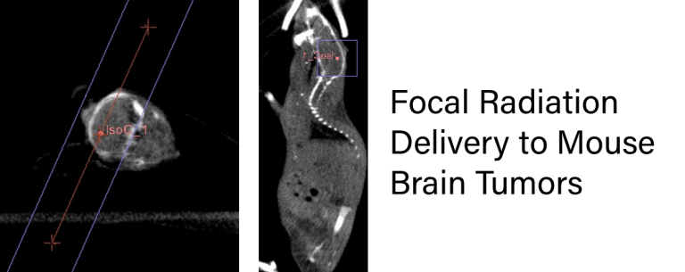
Great care is taken to spare surrounding tissue from the highest doses of radiation. By using an anatomical imaging technique such as CT, clinicians can create a treatment plan that delivers a precise therapeutic dose to the tumor alone.

We have joined with Xstrahl, a world leader in preclinical targeted x-ray irradiators, to extend this powerful technology to you as part of our service offerings. Xstrahl’s Small Animal Radiation Research Platform (SARRP) incorporates CT imaging with precise radiation delivery to enable researchers to pinpoint an exact anatomical target.

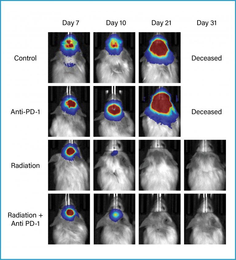
This small animal irradiator can then deliver single or multiple beams of radiation to the target with the utmost accuracy, matching the clinical techniques used in oncology departments around the world. This precision becomes important particularly in the evaluation of immune activating therapies where whole animal irradiation could inhibit and/or directly alter the animal’s innate immune system. The combination of precision radiation delivery and our expertise in immuno-oncology allows you to execute complex and powerful study designs.

Treatment of Orthotopic GL261-Luc Brain Tumors with Focal Radiation, Anti-PD-1 Antibody, or the Combination:

  • SARRP or anti-PD-1 antibody monotherapy provided improved anti-tumor activity over the control group

  • Treatment with SARRP and anti-PD-1 antibody in combination increased overall survival of either single agent treatment

  • The combination of precision radiation delivery and expertise in imaging and oncology pharmacology allows for the execution of complex and powerful study designs

Focal Radiation

  • Incorporation of CT imaging for precision radiation delivery

  • SARRP matches clinical techniques used in oncology departments around the world

  • Focal radiation can be used in SCID and NSG mice with less toxicity

  • Bilateral tumor studies can be run to evaluate potential abscopal effects

  • Focal radiation can be combined with immune modulating agents, chemotherapies, and many other therapeutic test agents

Contact us today to discuss the use of focal radiation for your next study.

Proof-of-Concept Studies

As a means to utilize the technology in a proof-of-concept experiment, mouse B-cell lymphoma tumor line A20 was implanted in the flank bilaterally in immune-competent mice. The right side tumor was specifically treated with a focused, fractionated dosing schedule of 2Gy five days a week for two cycles. On this dose schedule and duration, we began to see growth inhibition in the treated tumors. Contra-lateral tumors in the same mice were unaffected by the radiation treatment and matched vehicle control. This type of study design is easily modifiable to suit much higher radiation doses performed in a less frequent time frame as well. To validate this mouse glioblastoma cell line, GL-261-Luc was implanted intracranially into immune competent mice. Single high dose of either 10 or 15Gy was delivered specifically to the brain and bioluminescence imaging was used to monitor disease progression over time. We found that while a single dose of 10Gy resulted in 25% overall survival, a single dose of 15Gy resulted in 100% long term survival.

These types of datasets allow us to collaborate with our clients on developing specific study designs that meet their experimental needs.

Let's start a conversation

Contact Us